Is rheumatoid arthritis associated with extra-articular features and comorbidities?
Rheumatoid Arthritis (RA) is associated with a range of extra-articular features that manifest in several body systems and physiotherapists should be alert to these. These presentations are most commonly cutaneous (e.g. nodules) and neurological, but they can also seriously affect the cardiovascular and cardiopulmonary systems, and occur in up to 40% of people with RA. These manifestations are largely a result of chronic inflammation. People with RA also have increased risk of lymphoma.
Co-morbid health conditions may also occur in people with RA. They are sometimes a direct consequence of the disease (for example, increased risk of lymphoma, skin cancer and osteoporosis) and sometimes a consequence of drug therapies. For example, glucocorticoid (steroid) drugs may cause osteopenia and osteoporosis, while immunosuppressive agents (e.g. methotrexate) may increase susceptibility to infections and malignancies.
Practice points
- Early identification and early medical management decreases the risk of extra-articular features and comorbid conditions
- Physiotherapists regularly see patients’ skin and therefore should be particularly alert to skin cancers. Australia has the highest incidence of skin cancer in the world, and the incidence of skin cancer is greater in people with RA. The Cancer Council of Australia recommends that people at higher risk of skin cancer, e.g. people with RA, especially those on DMARDs or bDMARDs, have regular skin checks.
- Physiotherapists prescribe and oversee exercise programs for people with RA. Consequently, they may be the first to notice changes in a patient's cardiopulmonary capacity and exercise tolerance. These changes may indicate cardiopulmonary or cardiovascular impairments that should be investigated by a medical practitioner.
How do you recognise extra-articular features and co-morbidities associated with rheumatoid arthritis?
The key to identifying extra-articular features is listening and looking.
- Listen to the issues raised by a patient during the subjective examination.
- Are the presenting complaints unusual?
- Could presenting complaints that are non-musculoskeletal in nature be associated with RA?
- Does the patient also complain of malaise, weight loss, fatigue?
- Look for the signs of extra-articular features
For a summary of practice-relevant signs and symptoms of extra-articular features of RA, see the table below.
| Body system | Manifestation | What to look for |
|---|---|---|
| * severe manifestations † defined as chronic polyarthritis, neutropenia and splenomegaly. SOURCE: Briggs et al 2013 | ||
| Skin | Nodules Raynaud’s sign Major cutaneous vasculitis* |
Single or multiple subcutaneous nodules, >5mm diameter. Usually painless and on extensor surfaces. Colour change (paleness) of extremities fingers/toes in response to cold or stress Red or purple spotting on the skin, which does not blanch with pressure. Leg ulcers and peripheral gangrene |
| Pulmonary | Bronchiolitis obliterans organizing pneumonia (BOOP) Pleuritis* and pleural effusion Interstitial lung disease* |
Dry cough, dyspnoea, wheezing, crackles on auscultation Sharp chest pain with deep breathing, coughing, sneezing, laughing Dyspnoea, cough |
| Cardiovascular | Pericarditis* Vasculitis* Ischaemic Heart Disease |
Chest pain, dyspnoea. Palpitations. Signs of ischaemia or necrosis in affected organs/tissues. Reduced cardiac capacity. |
| Neurological | Mono neuritis multiplex* or peripheral neuropathy | Acute sensory and motor neuropathy in one or more nerve trunks, occurring as a result of vasculitis, compression or diabetes |
| Visual | Sjögren's syndrome Episcleritis or scleritis* |
Dry eyes and mouth. Skin, nose and vaginal dryness also present. 'red eye' - redness of the white part (sclera) of the eye, eye pain with possible radiation to the jaw, photophobia, decreased visual acuity |
| Haematological | Felty's syndrome*† | High rate of bacterial infections, fever, weight loss, fatigue. |
| Skeletal body system | Osteoporosis | Minimal trauma fracture, height loss. Be vigilant about bone fragility. |
Important
Morbidity and mortality associated with cardiovascular disease is one of the most common extra-articular manifestations related to RA. Physiotherapists should monitor cardiovascular status closely and work with patients to address risk factors for cardiovascular disease or on-refer if low confidence in this area. In particular, support to cease smoking is essential.Below are some examples of extra-articular manifestation of RA.
-

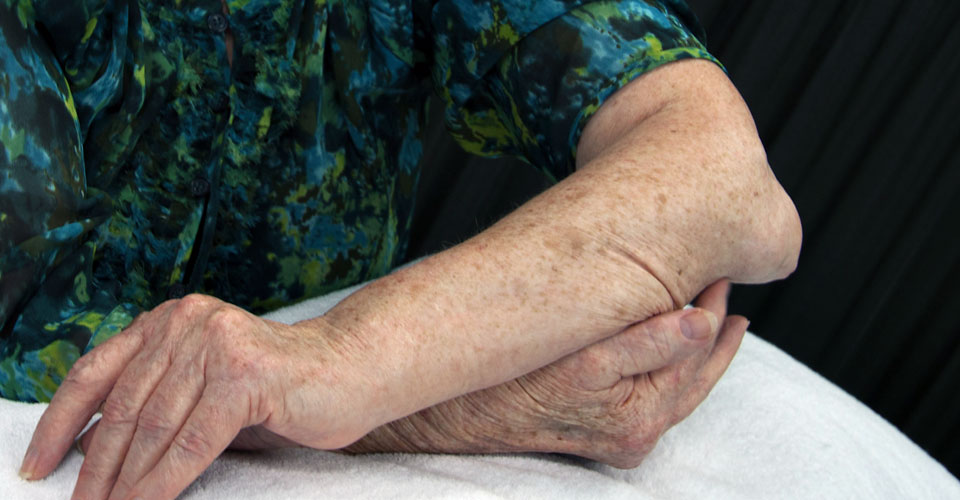
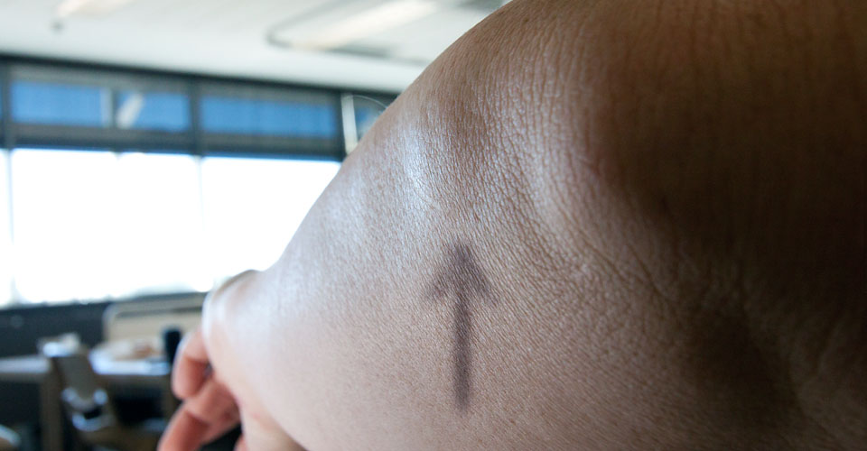
Nodule elbow
Subcutaneous nodule over left elbow.
-
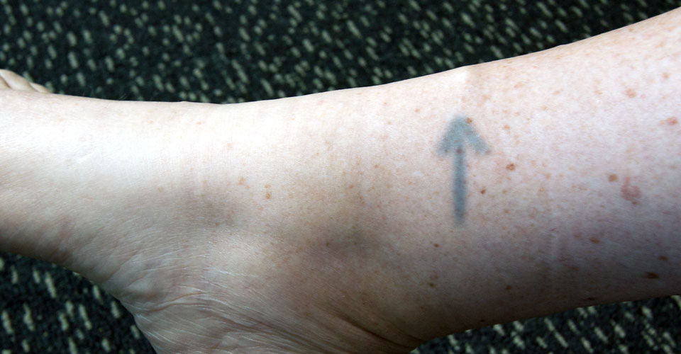
Nodule ankle
Subcutaneous nodule on anterior aspect of lower leg.
-
Nodule elbow
Subcutaneous nodule on ulnar side of forearm.
-
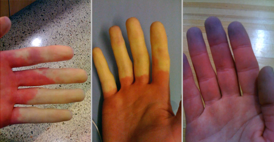
Raynaud’s sign
Examples of Raynaud’s syndrome.
-
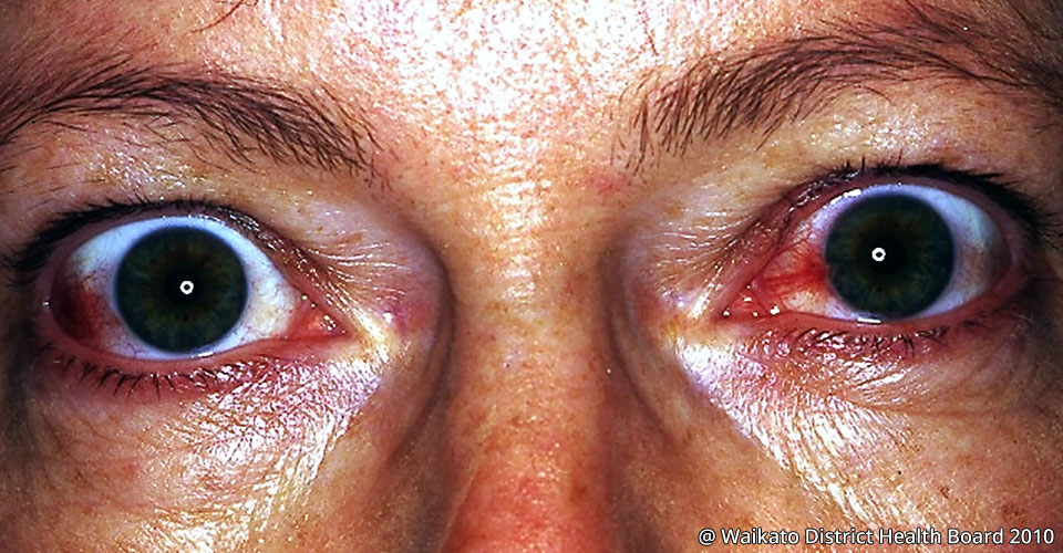
Episcleritis or scleritis
Example of RA associated episcleritis, termed ‘red eye’.


Recognising co-morbid conditions
Practice-relevant signs and symptoms of co-morbid conditions associated with RA may also present in practice. See the table below for common examples.
| Body system | Co-morbidities / complications | What to look for |
|---|---|---|
| * occurs as a consequence of subluxation of cervical spine joints. SOURCE: Briggs et al 2013 | ||
| Neurological | Cervical myelopathy* | Neck pain, upper limb pain, sensory and motor changes in upper limbs, gait disturbances. Be vigilant about the possibility for cervical spine instability (C1/2 specifically) |
| Metabolic / Endocrine | Osteoporosis Steroid-induced diabetes mellitus |
Minimal trauma fracture, height loss. Be vigilant about bone fragility. Painful peripheral neuropathy |
Below you will see some radiological images of cervical spinal instability.
-
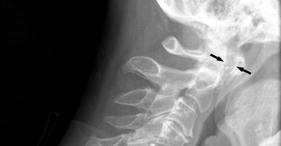
Cervical spine instability flexion
Plain radiograph of the cervical spine showing 5 mm of anterior subluxation (arrowed) of C1 on C2, reflected in an increased anterior atlanto-dens interval (AADI) on flexion.
-
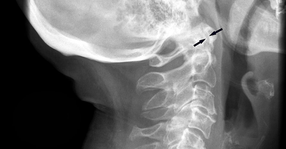
Cervical spine instability extension
While the anterior atlanto-dens interval (AADI) increases on flexion, it is reduced with cervical extension.
Practice points
- Identification of extra-articular features in practice often requires a thorough subjective assessment with screening questions for:
- body systems (e.g. dyspnoea, skin lesions, painful eyes)
- articular and peri-articular features
- Taking a thorough patient history and physical examination is critical in patients with RA.
- Cardiac, neurological and pulmonary manifestations have the potential to cause considerable disability for patients and influence on physiotherapy management. For example, reduced cardiac, pulmonary and neuromuscular performance may lead to fatigue, dyspnoea and reduced capacity to engage in aerobic and strengthening exercise and perform functional tasks.
- Physiotherapists should work with their patients to address modifiable risk factors for morbidity and mortality associated with cardiovascular disease, such as managing obesity and optimising nutrition, physical activity and lifestyle choices such as smoking and alcohol consumption.
- Cervical spine involvement in RA can take many forms. Reported findings include vertebral endplate erosions, spinous process erosions, and apophyseal joint changes, such as osteoporosis, blurring, and fusion. The most characteristic lesions are subluxations of the atlantoaxial complex (AAS), most commonly in the anterior direction (Zhang and Pope 2015). In a recent systematic review and meta-analysis of studies published between 1960-2014, Zhang and Pope reported a decrease in the prevalence of AAS over the last 50 years, most likely related to the introduction of DMARDs. However, anterior AAS was still reported with a prevalence of 27% and cervical myelopathy 5% in people with RA. The rate of new or progressive cervical myelopathies was calculated at rate of 1.5 per 100 patients with known cervical subluxations per year.
If I identify extra-articular features in practice, what should I do?
- Refer
- Modify management
- Educate
Refer
Refer to a medical practitioner when in doubt. Referral communications should highlight the findings that are suggestive of any extra-articular features.
Modify management
Physiotherapy management approaches may need adjustment to accommodate extra-articular manifestations and comorbid conditions relevant to each individual patient. You should:
- Consider exercise tolerance and goals for aerobic conditioning (please see our exercise evidence table for further information.)
- Consider implications and management of bone fragility from osteopenia/osteoporosis and cervical myelopathy
- Osteoporosis: encourage weight bearing exercise, balance exercise to address falls risk, postural education and exercise. Avoid strong manual therapy techniques in the context of severe osteoporosis
- Cervical myelopathy: consider cervical spine joint instability. Avoid strong manual therapy techniques in the upper cervical spine. On-refer if in doubt
Educate
About:
- Optimising lifestyle habits (smoking cessation, weight loss and relaxation techniques)
- Maintain strength and fitness
- Compliance with therapy and monitoring
Summary
- RA is more than a musculoskeletal condition. It is associated with extra-articular features in several body systems and these are potentially life-threatening
- The presence of extra-articular features usually indicates a more severe disease status and poorer prognosis. Therefore, physiotherapists needs to be able to identify such features early and refer to a medical practitioner